In my Anatomical Visualization class, the third assignment was illustrating an anomalous vein entering the heart taken from an article provided. The purpose of the assignment was to show volume of forms as well accurately portraying the various anomalies.
What’s so different you say?
Well the left hepatic vein is directly dumping into the right atrium, versus the inferior vena cava. The posterior vein of the left ventricle and middle cardiac vein are independently dumping in to right atrium as well, versus the coronary sinus. The coronary sinus is also a little unsual because of its size and placement, slightly left of entrance of the left hepatic vein. The ligamentum venosum also has it’s hand on the portal vein.
How and What do you use for references for this?
Booooooooooks, are where it’s at. They were valuable for this assignment to find placement of the heart on the liver, the relationships, as well as texture of tissues. Another wonderful source was our own cadavers! There were two hearts cut out, that were the chosen ones, to be saved from further dissection from our groups. In our lab we had a heart situated on a stand, held merely by a probe, and lamp brought in to create top left lighting. We had a lovely set up for sketching, but that was just for the heart. The liver was not as simple. The livers were left mostly intact in the bodies and finding other valuable resources weren’t as easy. Another one of our teachers had a model of a liver that we could use, useful so that you could physically hold the plastic (whatever material) liver, as get an understanding of it’s shape.
The point: Being able to take infomation that we know, and find solutions to represent the task, even though all that information we need isn’t sitting in front of us. We gotta use our brains man!
Oh yes, one more thing:
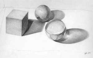
SHAPES! This task was given along with assignment to further our understanding of light on form. I made a still life. The highlight of this, my acrylic sphere, yes.

